341 An an ex radiographer from my memory and the remaining textboks I still have you would have to had smoked quite a lot for the effects to. A chest X-ray can be used to define abnormalities of the lungs such as excessive fluid fluid overload or pulmonary edema fluid around the lung pleural effusion pneumonia bronchitis asthma cysts and.

Chest X Ray Basic Interpretation X Ray Anatomy Radiology
The usual care group received standard medical care and no regular screening X-rays.

Chest x ray smoking. Routine screening for lung cancer with chest radiography or sputum cytology in asymptomatic persons has been abandoned because the yield of chest x-ray screening is low 04 cancer. Public health has emphasized the prevention or discontinuation of tobacco use. The patient is facing towards the left on the lateral view.
29th Nov 2002 1823 2 Cuddles. These scans use a small radiation dose 01millisievert or. It can help your healthcare provider see how well your lungs and heart are working.
The intervention group was offered annual screening chest X-rays for up to 4 years. A chest X-ray can also detect some abnormalities in the heart aorta and the bones of the thoracic area. Certain heart problems can cause changes in your lungs.
Include all chest x-rays performed within the audit time period for each patient group. Medical tests for smokers. Recently a new low dose ct imaging has been recommended for long term smokers that may identify lung cancer at an earlier stage than a chest x-ray.
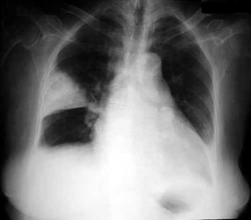
The classic finding on a smokers chest film is increased lung volume which in turn is caused by breakdown of lung tissue. Evidence from a population-based study in Italy springermedizinde Skip to main content. Look Out for Dirty Lungs in Chest X-Ray in Smokers.
Walking chair or trolley. Chest X-Ray is a type of X-Ray commonly used to detect abnormalities in the lungs. Researchers report that physical exams lung function tests and chest x-rays are not sensitive enough to pick up early damage from smoking.
A chest X-ray is an imaging test that uses X-rays to look at the structures and organs in your chest. Now most people would be definitely worried if you suggested that they have a chest x-ray every day for the rest of their lives. Often one can actually see holes in the lungs called bullae.
Hi moose5 smokings damage to your lungs is visible on an chest x-ray. How many cigarettes would one have to smoke untill it appeared on a chest x-ray. The groups were similar at the start.
Exclude portable and. Mode of transport of the patient eg. Others estimate that a cigarette addict may get exposed annually to a dose of radiation equivalent to 22000 chest x-ray examinations.
In the study 31 smokers who tested normal in every other way showed genetic changes in their lungs similar to those found in people with lung cancer. Each chest x-ray is checked whether it is an AP or PA using RIS and PACS. Thereafter use of nicotine chewing gum helps the patient to quit smoking.
But some of these people quite happily smoke sometimes up to two. S moking is a habit very difficult to get rid of. The historical observational studies of chest x-ray CXR screening for lung cancer LC showed improved LC-specific survival but no reduction of LC-specific mortality 1-6Four case-control studies performed in the 1990s in Japan on a population level however suggested CXR screening effectiveness as indicated by LC mortality reductions of about 40 78.
The PA exam is viewed as if the patient is standing in front of you with their right side on your left. ISZ - not the end of the world but you can see it from here. Characteristic changes consistent with pulmonary disease is evident with chronic smoking.
The question is how to motivate these smokers. It requires strong motivation. In this Episode we compa.
The diameter of the chest is increaed which is best seen on a view from the side and the diaphragm is pushed down and flattened. The time the chest x-ray was performed. The dose rate depends on the radioactive content of the tobacco the puff.
1 Show them statistics that smoking is the commonest cause of myocardial infarction and sudden death. About 45 had never smoked 42 were former smokers and 10 current smokers. The art and science of thoracic imaging.
2 Convince them that it is the. Chest X-ray Some doctors insist on yearly chest X-rays for all patients who smoke tobacco. Prospective trials have shown no evidence that screening reduces mortality.
Winters and DiFranza claimed that a person smoking 1 12 packs of cigarettes per day receives a dose to certain regions of the lung equal to 300 x-ray films of the chest per year. The films are read together. Chest X-Ray and smoking.
Is chest X-ray screening for lung cancer in smokers cost-effective. Radiographer who has taken the chest x-ray - this may be kept confidential. The standard chest examination consists of a PA posterioranterior and lateral chest x-ray.
Resources practice education and other topics on Chest X-Ray.

Pin On Thorax

Chest X Ray Of A 46 Year Old Patient Smoker With Pulmonary Langerhans Cell Histiocytosis Showing A R Radiology Langerhans Cell Histiocytosis Langerhans Cell

Pin On Rt

Wallpaper Backgrounds Get Abstract Desktop Wallpaper Backgrounds Skull Wallpaper Xray Art Posters Art Prints

Emphysema Xray What Are The X Ray Findings Of Emphysema Increased Radiolucency Black Along With Any Other Medical Radiography Radiology Imaging Radiology

Pancoast Tumour Radiology Case Radiopaedia Org Radiology Radiology Imaging Pulmonology

Pin On Why Smoking Is Bad For Smokers Lungs

Pin On What Smoking Does To You It Kills You
Sarcoidosis Is A Non Caseating Granulomatous Multi System Disease With A Wide Range Of Clinical And Radiographic Manif Radiology Imaging Human Body Icu Nursing

Pin On Critical Thinking About Poverty

Sarcoidosis There Is A Widespread Predominantly Reticulonodular Pattern With Relative Sparing Of The Lung Bases Ther Radiology Radiology Imaging Pulmonology

Chest X Ray Chest Radiography Nurse Study Guide

Interstitial Lung Disease Life Expectancy Prognosis Treatment Symptoms Causes Asthma Treatment Lung Disease Chronic Obstructive Pulmonary Disease
Pulmonary Infarction Wedge Infarct Radiology Medical Imaging Nuclear Medicine

Learningradiology X Ray Radiology Medical

Wedge Shaped Consolidations In Right Lung Middle Zone Could Be Infarct Associated Blunting Of Cardiophrenic And Costophre Radiology Langerhans Cell Thoracic

Pin On Anti Smoking Ads

Pin On Angie S Hypnosis To Quit Smoking

Right Upper Lobe Pneumonia Radiology Case Radiopaedia Org Radiology Radiology Imaging Pneumonia

